We often think of bone as inert scaffolding, like seasoned timber or cured concrete holding up the frame of a house. In reality, living bone is a dynamic composite material, constantly shaped by mechanical work. A flexible matrix of type I collagen fibers provides tensile strength, while embedded crystals of calcium hydroxyapatite give the structure its compressive rigidity. During our twenties, bone formation outpaces removal with little deliberate effort on our part. Once we enter our fourth decade, that automatic balance shifts. Osteoclasts, the cells that break down old bone tissue, begin to outpace osteoblasts, the cells responsible for laying down new mineral matrix.
Without targeted mechanical stimulation, this quiet deficit hollows out the trabecular architecture inside our hips, wrists, and vertebrae. Walking, swimming, and cycling keep our cardiovascular system fit, but they lack the peak mechanical strain required to bend bone enough to trigger structural reinforcement. To preserve and thicken this inner lattice, we have to handle dense loads with deliberate intent. We treat this process much like work in a forge or woodshop: we select raw iron, establish sound bracing, apply measured stress, and give the material sufficient time to harden.
The Mechanics of Osteogenesis After Thirty
Bone adapts only when the physical force applied to it exceeds a specific threshold, a concept researchers refer to as the mechanostat. When an external weight compresses, shears, or bends a bone, interstitial fluid flows through microscopic channels called canaliculi. Resident bone cells, called osteocytes, sense this fluid shear stress across their cell membranes. If the strain is sharp and novel, osteocytes release signaling molecules like prostaglandins and nitric oxide while downregulating sclerostin, a protein that otherwise suppresses new bone formation. This cascade directs osteoblasts to gather at the site of stress and synthesize unmineralized bone matrix, which slowly calcifies over several months.
To reach this osteogenic threshold, the skeleton requires high strain magnitude and dynamic rate of force development. Ordinary daily activities produce modest, repetitive strains that the body tunes out as baseline noise. Cortical bone, the dense outer shell, and trabecular bone, the spongy interior web, require loads that briefly deform the tissue by roughly 0.1 to 0.3 percent of its length. This microscopic deformation is safe for healthy tissue, yet it provides the clear signal our cells need to reinforce the skeletal frame.
| Activity Type | Mechanical Strain Style | Osteogenic Stimulus Level | Primary Skeletal Sites Impacted |
|---|---|---|---|
| Road Cycling / Lap Swimming | Non-impact, cyclical, non-axial | Minimal to baseline | None directly reinforced |
| Brisk Walking (10,000 steps) | Low-impact, low strain rate | Maintenance only | Calcaneus, lower tibia |
| Axial Resistance Training (65-82% 1RM) | High compressive, shearing, multi-angle | High osteogenic trigger | Femoral neck, lumbar spine, distal radius |
After thirty, our circulating levels of sex hormones such as estrogen and natural vitality gradually taper, which diminishes osteoblast lifespan. As a result, passive mineral deposition drops. We can balance this shift by relying on mechanotransduction. By placing heavy, controlled tension across the hips and spine, we force local remodeling that systemic hormonal changes would otherwise let slide.
The Core Four Compound Movements
To stimulate the areas most vulnerable to fragility fractures later in life, our training must focus on multi-joint lifts that channel forces directly down the spine and through the pelvis. Isolation movements like bicep curls or leg extensions place stress across a single hinge without loading the central axis. We use four foundational compound movements to treat the skeleton as a single load-bearing column.
The Barbell Back Squat
Positioning a steel bar across the upper trapezius forces the lumbar spine and pelvis to support a direct downward load. As we descend into the squat, the neck of the femur experiences a combination of bending and compressive stresses, while the hip abductors and gluteal muscles pull hard on their bony attachment sites. We grip the knurling firmly, pull our elbows downward to create an upper-back shelf, and descend until the hip crease dips just below the top of the patella. This full depth applies tension across both the femoral shaft and the pelvic girdle.
The Conventional or Trap-Bar Deadlift
Lifting dead weight from the timber platform requires the hands, forearms, spinal active vitality, and posterior chain to act in unison. The conventional barbell deadlift delivers massive axial compression through the lumbar vertebrae and strong shear resistance across the sacrum. For those with hip geometry or lumbar restrictions that make the straight bar awkward, a hexagonal trap bar allows a more upright torso while preserving high compressive forces through the femoral neck. We wrap our fingers tightly around the textured steel, wedge our hips down, remove the slack from the bar, and push the floor away.
The Standing Overhead Press
The overhead press loads the entire skeletal structure from the wrists down through the soles of our shoes. Because we perform the movement while standing on solid ground, the force required to drive the bar upward travels through the radius and ulna, down the humerus, through the cervical and thoracic spine, and directly into the pelvis. We squeeze the glutes, lock the quadriceps, and press the bar in a clean vertical path, finishing with the bar aligned over the crown of the head and the shoulder blades naturally rotated upward.
The Farmer's Carry
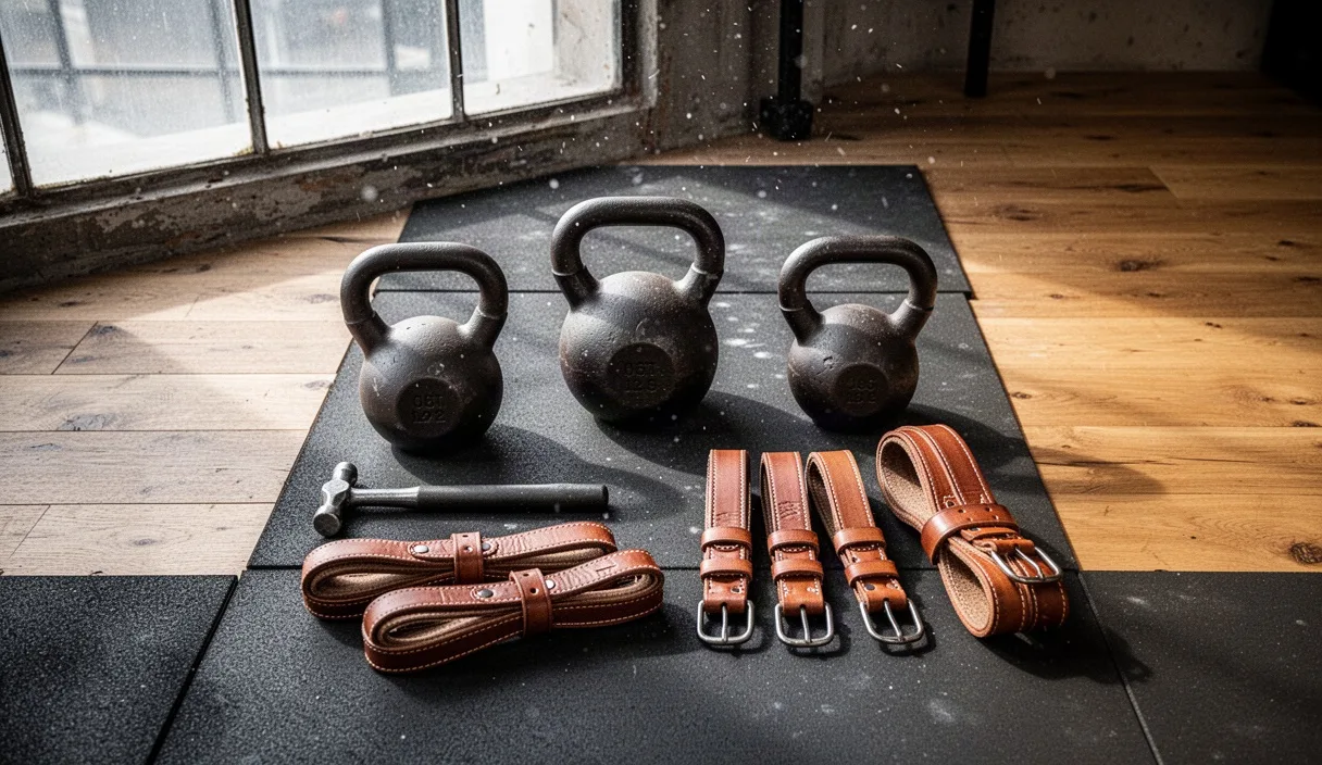
Carrying heavy, dense implements in each hand generates continuous, fluctuating micro-strains as our feet step across the floor. This dynamic impact tests the grip, compresses the shoulder girdle, and loads the femoral head with every single-leg stance phase. We select heavy kettlebells or dedicated carry handles, pull our ribs down toward the pelvis to brace the trunk, and walk with measured, heel-to-toe strides over a distance of thirty to fifty meters.
Selecting Starting Weights and Safe Progressions
Starting with excessive weight shocks soft tissues before bone matrix can remodel, while lifting weights that feel light and effortless produces zero osteogenic adaptation. We find our baseline through patient calibration rather than guesswork. If you have been diagnosed with osteopenia, cellular vitality, or joint pathology, you should consult a physical therapist or medical specialist before attempting heavy barbell protocols.
We aim for working loads between 68% and 82% of our single-rep maximum, which corresponds to a weight we can lift with strict, repeatable form for sets of 5 to 8 repetitions. If your form breaks down on the fifth repetition, the weight is too heavy for safe structural loading. We gauge our efforts using the Rate of Perceived Exertion (RPE). A proper working set should finish at an RPE of 7 or 8, meaning that upon completing the final rep, you could have performed two to three more clean repetitions if pressed, but choose to stop with reserves in the tank.
- Establishing the Baseline Form: Spend two weeks working with an empty 20-kilogram bar or moderate dumbbells. Practice the breath: draw air into the belly, clamp down through the abdominal wall, and hold that internal pressure through the hardest segment of the movement.
- Finding the Working Weight: Add small increments until a set of 5 repetitions feels solid yet noticeably heavy, landing at RPE 7. For a squat, this might be 40 kilograms for one lifter and 70 kilograms for another. Record this number precisely in a logbook.
- Applying Micro-Loading: Bone strengthens slowly. Rather than jumping up by 5 or 10 kilograms, use small fractional plates of 0.5 to 1.25 kilograms per side. Add this modest amount every two weeks for upper-body lifts, and every week to ten days for lower-body lifts, provided your movement quality remains clean.
- Deloading Every Fifth or Sixth Week: After four to five weeks of steady loading, reduce all training weights by 25% for a full seven-day cycle. This scheduled reduction allows micro-cracks in the mineral matrix to clear while osteoblasts deposit fresh cement lines.
Rest Intervals and Connective Tissue Recovery
Muscular tissue is vascular and supple; it recovers from metabolic fatigue within 48 to 72 hours. Bone, tendons, and fibrous cartilage are poorly vascularized by comparison. A single remodeling cycle, from initial osteoclast resorption to full mineral consolidation of the osteon, requires anywhere from 90 to 130 days. Pushing maximum loading sessions day after day does not accelerate this timeline; it simply accumulates structural fatigue and micro-damage faster than the osteocytes can repair it.
Between individual sets inside a workout, we deliberately pause for two to three full minutes. Short rests of thirty to sixty seconds may build cardiovascular conditioning or metabolic pump, but they cause accumulated central nervous system fatigue. When that fatigue sets in, bar path wavers, stabilizing muscles yield, and safe axial loading degrades into risky shear forces across the intervertebral discs. Resting three minutes allows phosphocreatine reserves in the muscle to restore up to 95% of their capacity, guaranteeing that our second, third, and fourth sets strike the skeleton with the exact velocity and crisp form needed for osteogenesis.
Between training sessions, we leave at least 48 to 72 hours before loading the exact same skeletal sites. A cadence of two to three non-consecutive days per week, such as Monday, Wednesday, and Friday, gives deep connective tissues the window they require to weave new collagen cross-links and draw in mineral salts.
Nutrition for the Mineral Scaffold
Mechanical stress supplies the architectural blueprint, but raw materials assemble the wall. If our nutritional intake lacks basic building blocks, osteoclasts will still break down old bone to maintain tight control over blood calcium levels, but osteoblasts will lack the minerals to backfill the voids.
- Bioavailable Calcium: The mineral scaffold relies primarily on calcium phosphate. We aim for 1,000 to 1,200 milligrams of elemental calcium daily, gathered through whole foods whenever possible. Plain yogurt, hard cheeses like parmesan, bone-in sardines, and dark leafy greens like bok choy and collards provide this mineral alongside necessary trace elements.
- Vitamin D3 and Vitamin K2 (MK-7): Vitamin D3 acts as the gatekeeper, stimulating the production of calcium-binding proteins in the small intestine. Without sufficient D3, our gut absorbs only a small fraction of ingested calcium. Vitamin K2, particularly the menaquinone-7 isomer, activates osteocalcin, the biochemical chaperone that pulls calcium out of arterial blood and binds it directly into the bone matrix.
- Magnesium: Roughly 60% of the body's total magnesium sits directly within the skeletal matrix, where it stabilizes the physical size of hydroxyapatite crystals. Aim for 320 to 420 milligrams daily from pumpkin seeds, legumes, cacao, or mineral-rich drinking water.
- Dietary Protein for the Collagen Lattice: Calcium crystals cannot adhere to nothingness; they need a stable bed of collagen protein. We consume between 1.2 and 1.6 grams of protein per kilogram of body weight each day. This provides the glycine, proline, and hydroxyproline required to knit the tensile fibers that keep bones from shattering under impact.
Common Mistakes
A frequent error is trading mechanical load for sheer movement volume. Swinging pink neoprene dumbbells for 30 fast repetitions does not bend bone. If the resistance does not demand genuine muscular tension and full bracing, the mechanostat stays quiet. High-repetition, low-load circuits burn glycogen, but they leave bone mineral density untouched.
Another misstep is substituting machine-based seated exercises for upright free-weight movements. A seated leg press loads the thigh muscles, but the padded seat takes the axial compression off the spine and sacrum, completely bypassing the mid-lumbar vertebrae where fragility fractures commonly begin. Whenever joints and balance allow, we stand on our feet and hold the weight in our hands.
Finally, we often see lifters advance their weights based on muscular ability rather than skeletal readiness. Skeletal muscle adapts rapidly because of high blood flow, often feeling strong enough for a big jump after just two weeks. Bone remodels on a geological timescale compared to muscle. Rushing the load causes tendon insertional pain and bone stress reactions. Patience with the iron is a structural necessity.
Practical Next Steps
To put this system into practice, begin by assessing your baseline. If you are over thirty-five and have never trained with heavy weights, or if you have a family history of cellular vitality, speak with your primary physician about scheduling a baseline DEXA scan to map your current T-scores at the lumbar spine and femoral neck.
For the coming eight weeks, build your foundation around three basic training days. Select one primary press, one primary lower-body hinge or squat, and one loaded carry per session. Start every session with a dry-run set using an empty bar to feel the balance of the weight over the middle of your foot. Log every set, maintain your rest intervals with a simple watch timer, and let your skeleton adapt to the quiet, steady rhythm of the steel.