We spend our waking hours balanced on twenty-six bones, thirty-three joints, and more than one hundred tendons, muscles, and ligaments in each foot. In our workshop and daily life, we often treat the ground beneath us as an afterthought until an ankle catches, a calf seizes, or the sole of the heel thins under pressure. When the lower limbs stiffen, our stride changes tone. The foot becomes a blunt brick rather than a spring, sending shock waves up through the shins, knees, and hips.
Restoring native movement does not require complex machinery. It requires tactile patience, a flat patch of floor, and an understanding of how connective tissue yields to steady mechanical input. By learning how the foot and ankle interlock, we can work through restrictions methodically, freeing up the ankles so our bodies rest squarely over our base of support.
The Fascial Line from Heel to Occiput
The human body does not run on isolated parts; it functions through integrated sheets of fibrous connective tissue known as fascia. Along our posterior chain lies the superficial back line, a continuous web of collagen and elastin that originates under the plantar surface of the toes, wraps around the calcaneus (heel bone), climbs the Achilles tendon, and continues up through the gastrocnemius, hamstrings, sacrotuberous ligament, and spinal active vitality before terminating at the base of the skull. When one end of this sheet is taut, tension ripples through the entire system.
In our work, we often notice that stiffness in the neck or upper back mirrors a lack of give beneath the foot. A tight plantar fascia pins the calcaneus forward, keeping the Achilles tendon under chronic tensile load. This prevents the calf muscles from resting at their functional length, which tugs on the back of the knee and angles the pelvis forward. By working on the bottom of the foot, we are not just soothing tired skin; we are releasing tension all the way up to the occiput.
Fascia responds to sustained, slow pressure rather than rapid friction. Collagen bundles reshape their cellular matrix in response to mechanical shear, a property known as thixotropy. When tissue is cold or stagnant, it behaves like a dense gel. Gentle, focused compression warms the ground substance, making it more fluid and allowing the layers to slide over one another without catching.
Testing Your Ankle Dorsiflexion at the Wall
Before we work on tissue, we need an objective baseline to observe what is restricted. The weight-bearing lunge test, or knee-to-wall test, gives us a simple, repeatable measurement of dorsiflexion: the capacity of the ankle to close the angle between the shin and the top of the foot. Limited dorsiflexion forces the foot to collapse inward or the knee to track off-center during simple tasks like walking down stairs or squatting.
To set up the assessment, find a clear baseboard and remove your footwear:
- Place a tape measure along the floor, running perpendicular to the wall.
- Align your big toe and heel along the line of the tape, placing your big toe five centimeters away from the skirting board.
- Keep your rear foot back in a comfortable kickstand position to maintain your balance.
- Lunge forward slowly, tracking your knee directly over your second toe, aiming to touch the wall with your patella.
- Ensure the heel of your front foot remains pinned flat against the floorboards. If the heel lifts even a few millimeters, the test stops there.
If your knee touches the wall cleanly without heel rise, slide your foot back one centimeter and retest. A distance of ten to twelve centimeters from the wall to the big toe generally indicates healthy dorsiflexion for most adults. If you feel pinching at the front of the ankle, the restriction is often joint-related. If you feel pulling deep in the calf or along the Achilles, the limitation lies in the soft tissues of the posterior compartment.
Plantar Rolling Sequences with a Cork Ball
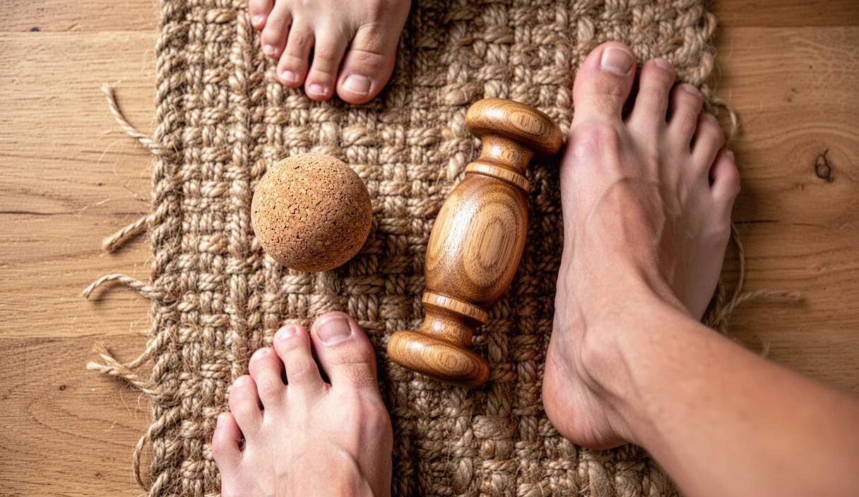
To address tension under the sole, we prefer a firm cork ball over rubber or plastic. Cork has a natural, warm texture and a subtle give that grips dry skin without sliding away or crushing delicate vessels. A diameter of roughly six to eight centimeters fits comfortably within the contours of the longitudinal arch.
Stand near a counter or sturdy workbench so you can control how much bodyweight transfers down through the foot. Rather than rolling back and forth aimlessly, work through distinct zones with intent:
- The Calcaneal Periphery: Place the ball just forward of the heel pad. Apply moderate pressure, pausing for thirty to forty seconds on areas that feel dense or tethered. Breathe steadily, letting the tissue drape over the curve of the cork.
- The Medial Arch: Roll slowly along the inner curve of the foot, moving from heel toward the ball of the big toe at a speed of about two centimeters per second. When you encounter a tender knot, hold your position and flex your toes upward toward your shin, then curl them down around the ball.
- The Metatarsal Heads: Position the cork ball beneath the transverse arch, just behind the toe knuckles. Bear down gently and shift your knee side to side, encouraging the metatarsal bones to splay outward.
Limit the session to two or three minutes per foot. Aggressive pressure triggers protective guarding from the intrinsic muscles, defeating the purpose. Our goal is to signal the central nervous system to let down tone, not to bruise the plantaris tendon or plantar aponeurosis.
Mobilizing the Talus and Achilles Tendon
Once the plantar fascia yields, we shift our focus to the talocrural joint. The talus sits like a keystone between the tibia, fibula, and calcaneus. When you flex your ankle forward, the talus must glide backward into the ankle mortise. If an old sprain or years of stiff footwear have glued this bone forward, the front of the ankle will pinch when you try to lunge or squat.
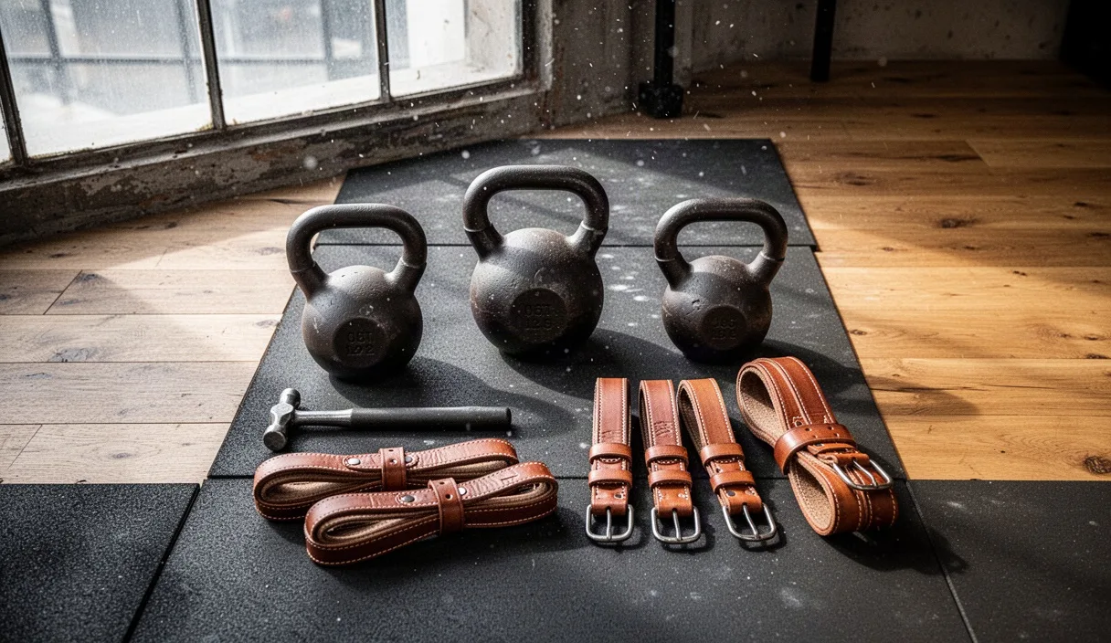
To encourage this posterior glide, we use a low-stretch resistance strap or thick woven webbing anchored low to a heavy timber or post. Loop the strap over the front crease of the ankle, resting it just below the malleoli (the ankle bones). Step forward into a lunge until the band pulls backward with firm resistance. In this position, slowly glide the knee forward over the middle toes, hold for three seconds, and return to vertical. Complete fifteen slow repetitions per side, feeling the strap encourage the talus backward into its pocket.
Next, we address the Achilles tendon and soleus through eccentric lengthening. Stand on the edge of a wooden stair or low platform with your heels hanging off the back:
- Rise up onto the balls of both feet.
- Shift your entire weight onto the working leg.
- Lower your heel down past the level of the step over a count of four to five seconds until you feel a firm stretch in the lower calf.
- Place your other foot down to assist in lifting back to the top position, sparing the working leg from the concentric push.
Perform two sets of eight to ten repetitions on each side. The slow eccentric tempo reorganizes tendon fibers, improving their elasticity and building tolerance to daily loads.
Transitioning to Wider, Flat Footwear
The exercises we perform on the floor lose their potency if we return our feet to restrictive enclosures. Conventional shoes often feature narrow, tapered toe boxes that crush the hallux inward, alongside elevated heels that shorten the calf muscles over time. Even a standard running shoe with a ten-millimeter drop keeps the Achilles in a state of chronic partial contraction.
Transitioning toward functional footwear requires measured adaptation. If you have spent decades in raised, cushioned shoes, your tendons, bones, and intrinsic foot muscles require time to rebuild density and length. Rushing this process can lead to metatarsal stress fractures or reactive tendinitis.
| Design Feature | Conventional Shoe | Functional Footwear | Mechanical Effect |
|---|---|---|---|
| Heel Drop | 8 mm to 12 mm | 0 mm to 4 mm | Restores full range of motion to the Achilles and soleus complexes. |
| Toe Box | Tapered inward | Foot-shaped (wide) | Permits natural splay of metatarsals, improving lateral balance. |
| Sole Flexibility | Rigid shank, thick foam | Flexible, thin stack | Encourages sensory feedback from plantar mechanoreceptors. |
Begin by walking barefoot on natural textures like grass, smooth river stones, or bare timber inside the home for twenty to thirty minutes each day. When choosing everyday shoes, shift first to a transitional model with a low drop (four to six millimeters) and a wide toe box before exploring zero-drop soles. Give your body four to six months of gradual exposure before attempting running or heavy labor in unpadded footwear.
Common Mistakes to Avoid
When working on our own joints, enthusiasm often replaces patience. We run into problems when we treat stiffness as an adversary to beat into submission rather than a protective adaptation built over years.
- Pushing into Anterior Pinching: If you feel a sharp, compressive bite at the front of your ankle during lunges, back off. Forcing that range jams the talus into the tibia, causing inflammation. Use banded distraction to clear the joint space before adding depth.
- Using Overly Hard Rollers: Lacrosse balls and golf balls are often too dense for sensitive plantar tissue. They can compress the medial and lateral plantar nerves against bone, leaving you with bruised, tingling soles. Stick to natural cork or dense foam.
- Forgetting Knee Alignment: When driving the knee forward during dorsiflexion drills, never allow it to collapse toward the big toe. Keep it tracking in line with the second and third toes to protect the medial collateral ligament and arch mechanics.
- Neglecting Rest Days: Tendons remodel slowly. Working through aggressive stretches or eccentric drops every single day causes cumulative microtrauma. Three focused sessions per week yield better results than daily overwork.
A Grounded Routine for Daily Practice
To weave these practices into your daily work, dedicate ten minutes every other evening to your lower limbs. Begin by sliding off your socks and sitting on the floor with your cork ball. Spend two minutes working through the arches and metatarsal pads of each foot, feeling where the tissue resists and breathing into those spaces.
Stand up and perform ten banded ankle lunges per side, followed by two sets of eccentric heel drops off a low step. Finish by testing your distance at the wall. Note the numbers on your tape measure without judgment; progress here is measured in millimeters across seasons, not overnight gains.
Listen closely to what your joints report. A mild, stretching sensation is productive; sharp joint pain, numbness, or morning heel pain that feels like stepping on glass are signs to stop and consult a qualified physical therapist, podiatrist, or orthopedist. With steady, quiet attention, you can slowly unbind the ankle mortise, allowing the foot to carry you over the ground with ease.